
TPLO Radiograph Positioning
Lateral View:
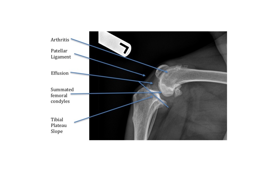
Obtaining appropriate stifle radiographs is imperative in the diagnostic work-up of a cruciate ligament injury. The lateral view allows us to evaluate for effusion and arthritis secondary to a cranial cruciate ligament injury. If the image is overexposed and you can’t see the patellar ligament, then you can’t evaluate for effusion.
The 90/90 lateral view (90° flexed at the stifle / 90° flexed at the hock) is required to accurately measure the tibial plateau slope for proper implant placement.
Summation of other structures (other leg, abdomen, etc.) over the stifle should be avoided.

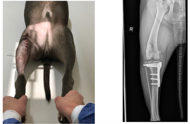
Appropriate positioning for pre- and post-operative films
Lateral and craniocaudal views at preoperative, immediate post-operative and 8 week recheck time points are shown. Notice on the lateral the tibia is parallel to the vertical cross-hair line and the femoral condyles are summated, ensuring no limb rotation. This allows full evaluation for degenerative disease and bone healing post-operatively.
On the craniocaudal view the limb should be pulled straight and rotated so the patella is centered. Sedation may be required to obtain appropriate views.